
Science
Benefits of Microscopy for the Use of Research Experimentation
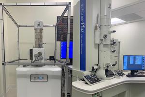
In the rapidly expanding field of microscopy, the scanning electron microscope (SEM) and transmission electron microscope (TEM) are at the forefront of ongoing research projects. Similar to the light source used in a standard compound microscope, the TEM uses transmitted electrons to produce a highly magnified image of a sample specimen. Similarly, the SEM provides detailed images of a sample specimen by bombarding the sample in a raster pattern using a focused electron beam.
There are major advantages to both the SEM and TEM, respectively. The SEM is mainly used to provide detailed imaging of the sample’s surface. Additionally, when used in conjunction with the EDS, or Energy Dispersive X-Ray Spectroscopy, the SEM is able to characterize the elemental composition of the sample. While the SEM focuses on the surface morphology of samples, the TEM focuses on thin slices of embedded specimen or a single layer of particles and can reveal more characteristics of the sample, including internal morphology and elemental composition, using the EDS.
These two powerful microscopes are available at American University for both research students and professors alike. Additionally, there are a number of research projects being performed on these two microscopes. Professor Shouzong Zou, chair of the Department of Chemistry, has ongoing research involving the shape-controlled synthesis of gold nanoparticles. As suggested by its name, the gold nanoparticles are too small to be observed with a normal compound light microscope. In other words, the SEM is first used as a screening tool to detect the presence of different shapes of gold nanoparticles, including cubic, octahedral, and rhombic dodecahedral. The TEM will then optimize the process of detection through high-resolution imaging. Eventually, these gold particles will be placed at the end of carbon nanofibers to increase the sensitivity of dopamine detection. The purpose of increasing the sensitivity of neurotransmitters is to potentially be able to use carbon nanofibers to detect neurodegenerative diseases at the earliest stage.
While this is only one application of the potential benefits of SEM and TEM analysis, the addition of these two instruments on campus over the past year have helped student researchers to learn more about their samples. Additionally, they have provided interesting opportunities for faculty researchers from neighboring universities.
These opportunities have excited those on campus and in particular, Andrea Brothers. According to Brothers, the instruments coordinator in AU’s Department of Chemistry, the microscopes on campus are beginner-friendly and can be used to study material from a number of different fields. Brothers joined the American University staff only a few months after the addition of both the microscopes on campus. She has been in charge of teaching and familiarizing student researchers with the complex sample preparation and discovery of samples at a nano-scale.
Due to the demanding sample requirements of the SEM and TEM, the sample preparation involved is complex and can be the most important step of the analysis. Normally, the SEM involves coating the sample with a metal substance such as gold. On the other hand, the TEM requires samples to be cut or prepared extremely thin, ranging from 40 to 90 nanometers. The SEM and TEM present an exciting opportunity for sample analyzation on a very small scale for all who are interested. Brothers has said that the TEM can be used to characterize tissues of organs and the organelles of individual cells.
As student researchers begin to work more with the SEM and TEM, there will be a production of different types of data. As we explore more applications at American University, their impact around the globe has already benefited a number of industries. A tangible application can be seen at nearby George Mason University, where researchers working with Ceres Nanoscience have worked to develop a non-invasive novel test to diagnose Lyme disease. More specifically, they have used both the SEM and TEM for the Ceres’ Nanotrap Lyme Antigen Test, in which the Lyme antigens from urine samples are concentrated and detected to reveal detailed information about the presence of the Lyme antigen in various stages of infection. While we at American University are only beginning our journey with these instruments, a lot of problems will be solved using the SEM and TEM.
While these two instruments, and especially the TEM, provide the campus with an ability to perform at a very high standard for most of its research, the number of advancements in the field of electron microscopy is constantly growing. There are researchers working to improve the resolution of both microscopes and increase the resolving power to smaller and smaller dimensions.